A Portal Triad Includes Which of the Following Anatomical Structures
Just so which structures are part of the portal triad. Each portal triad contains neural and lymphatic elements.

Liver Basicmedical Key
Textbook solution for Fundamentals of Anatomy Physiology 11th Edition 11th Edition Frederic H.
. It is a tube like anatomical structure formed by the convergence of the common hepatic duct and the cystic duct of the gallbladder. Branches of the hepatic artery carry oxygenated blood to the hepatocytes while branches of the portal vein carry blood with nutrients from the small intestine. It consists of the following five structures.
Which of the following statements correctly characterizes a portal triad. Each portal triad contains a branch of the portal vein hepatic artery and common hepatic duct. Intraoperative exsanguination is the primary cause of death and hemorrhage control should be the first priority.
The mechanism of this injury occurs when a lateral force to the knee is received while the foot is fixed on the ground. Portal triads are composed of three major tubes. Ducts right and left hepatic duct branches A.
Branches of the hepatic duct cystic duct and central vein. 1 pts Common bile duct Common hepatic artery Hepatic portal vein Hepatoduodenal ligament Hepatogastric ligament Proper hepatic artery The great saphenous vein drains into which blood vessel. Given that the portal vein carries mostly deoxygenated blood.
Solution for The portal triad found at each corner of a iver lobule consists of the following 3 structures choose 3 options. Martini Chapter 24 Problem 23CP. Proper hepatic artery an arteriole branch of the hepatic artery that supplies oxygen.
Anterior cruciate ligament ACL medial meniscus and tibial medial collateral ligament. Each portal triad contains a branch of the hepatic vein hepatic artery and common bile duct. It supplies oxygenated blood to the hepatocytes.
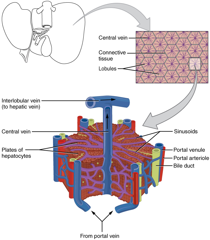
Injuries to the anatomical structures of the portal triad are rare and often lethal. The liver lobule is a hexagonal shaped structure. Vein portal vein E.
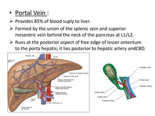
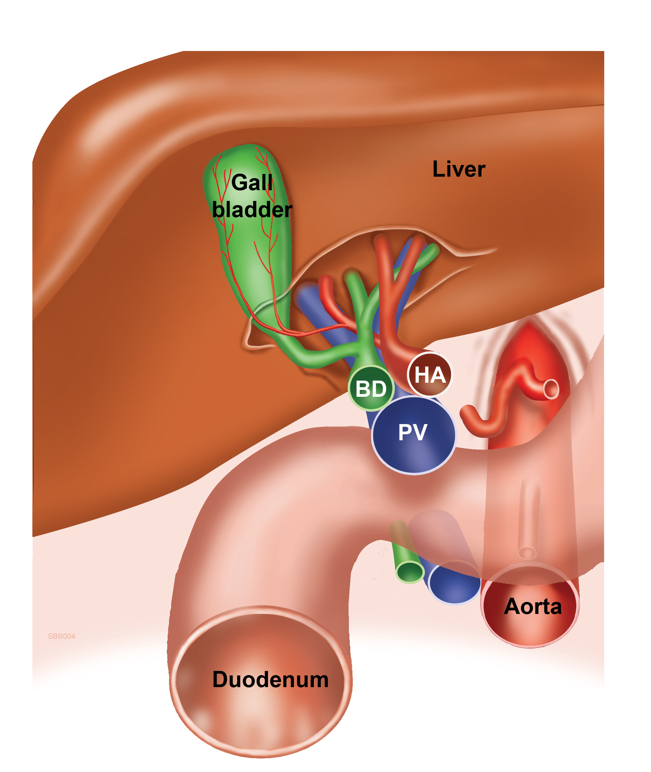
At the periphery of the hexagon are three structures collectively known as the portal triad. A useful mnemonic to help remember the order of structures in the porta hepatis from anterior to posterior is. The portal triad is formed by the portal vein PV the PHA and the common bile duct.
Bile duct injuries should be identified by intraoperative cholangiography and. A and C E. Hepatic portal vein a venule branch of the portal vein with blood rich in nutrients but low in oxygen.
The unhappy triad also known as a blown knee refers to a sprain injury which involves 3 structures present in the knee jointThese structures include. Asked Aug 27 2019 in Anatomy Physiology by amenahnomani. Melinda and Alex are co-leads on a project at work but their working styles differ greatly and they are having trouble agreeing on procedures and objectives.
What structures form the portal triad. The portal vein appears as the dot at the base exclamation point formed in this longitudinal view. Elena a member of their team who is relatively new to the company steps in and pulls together a crucial part of the project to meet a.
Each anatomical lobule is hexagonal-shaped and is drained by a central vein. Bile duct injuries should be identified by intraoperative cholangiography and repaired primarily or by enteric anastomosis. The portal arteriole is the branch of the hepatic artery.
What structures comprise a portal triad. Branches of the hepatic artery hepatic vein and hepatic duct. Intraoperative exsanguination is the primary cause of death and hemorrhage control should be the first priority.
Venule a branch of the hepatic portal vein entering the liver. Injuries to the anatomical structures of the portal triad are rare and often lethal. Branches of the hepatic artery hepatic portal vein and.
Which of the following statements regarding sonographic characteristics of hepatobiliary structures is correct. One or two small bile ductules of cuboidal epithelium branches of the bile conducting system. Bile canalicul Bile ductule Branch of hepatic artery proper Central vein Hepatic triad Branch of.
The hepatic vein hepatic artery portal vein and bile ducts all have bright echogenic walls. Arteries right and left hepatic artery branches V. At the angles of the liver lobule are portal triads.
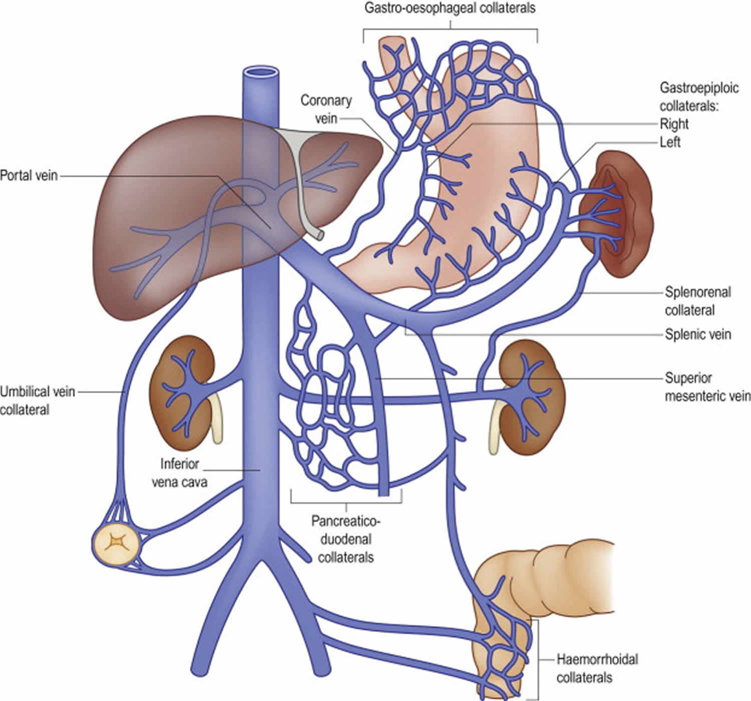
The lengthy axis is an imaginary line between two nearby central capillaries. The PV is formed by the unification of the superior mesenteric and the splenic vein. The bile duct carries bile products away from the hepatocytes to the larger ducts and gall bladder.
The point where the two ducts join is known as the ampulla of vater and is surrounded by a. Branches of the cystic duct central vein and hepatic artery. The brief axis is stood for by a shared boundary in between 2 nearby lobules along with the portal canals.
Anatomy and Physiology Correctly label the following microscopic anatomy of the liver. The portal vein and common bile duct share similar color-Doppler flow characteristics. With subtle fanning and adjustments of the probe the remaining structures of the portal triad the hepatic artery and common bile duct can be brought into view.
The common bile duct is the third and final structure in the portal triad. Arteriole a branch of the hepatic artery entering the liver. Portal Triad Injuries.
The portal triad consists of three vessels namely the portal arteriole portal venule and the bile duct. The axis of the traditional or physiological lobule is the main vein which is the beginning of the hepatic blood vessel. We have step-by-step solutions for your textbooks written by Bartleby experts.
It is usually found posterior to the proper hepatic artery and the common bile duct CBD within the hepatoduodenal ligament as part of extra hepatic portal triad. The pancreatic duct also merges with it as it nears the pancreas.
Accessory Organs In Digestion The Liver Pancreas And Gallbladder Anatomy And Physiology

Portal Triads Of The Liver Portal Triads Are Composed Of A I Portal Download Scientific Diagram

Anatomy Of Hepatobiliary Hepar The Largest Gland Of

Week 1 Biliary And Liver Flashcards Quizlet
![]()
Hepatoduodenal Ligament Anatomy And Contents Kenhub
Radiological Anatomy Of Hepatobiliary System

Liver
2

Southeast European Medical Forum Ppt Download

A Exploded View Of The Liver Demonstrating The Distribution Of Download Scientific Diagram
Cartoon To Remember The Position Of The Structures In The Hepatoduodenal Ligament English Labels Anatomytool

Liver Anatomy A Entire Organ And Blood Supply Blue Indicates Venous Download Scientific Diagram
Portal Vein Anatomy Function Embolization Thrombosis Hypertension

Histological Section Of The Liver In Mallard Duck Showing The Portal Download Scientific Diagram

Hepatic Lobule Histo Anatomiya

Gallbladder And The Biliary System Radiology Key

Copyright C The Mcgraw Hill Companies Chapter 16 Organs Associated With The Digestive Tract Organs Associated With The Digestive Tract Introduction The Organs Associated With The Digestive Tract Include The Salivary Glands The Pancreas The Liver And

Free Quiz Exercise Internal Anatomy Of Liver Internal Anatomy Of Liver Hepatic Vein Central Vein System Aorta Anatomy Liver Anatomy Portal System

2 14 Liver And Gallbladder High Yields Flashcards Quizlet


Comments
Post a Comment